What Is The Ideal Treatment For Heel Spur

Inferior Calcaneal Spur

Overview

While the term heel spur may create the impression of a sharp bony projection on the bottom of the heel that pokes the bottom of our foot causing our pain. Painful heel spurs are actually a result of damage to the soft tissue at the bottom of the foot. While this may be confusing, we’ll try to explain. Heel spurs is the more common name for a condition that is medically referred to as plantar fascitiis or heel spur syndrome. Plantar fasciitis is a location oriented term that refers to the bottom of the foot(i.e. plantar warts). Fascia is a tough, inelastic band. ‘itis’is a term used to describe something that is inflamed (i.e. tendonitis, bursitis).

Causes

One of the most common causes for the development of heel spurs is the wearing of shoes that are too tight. That?s why more women suffer from heel spurs more than men. Athletes who tend to stress their feet a lot, people are overweight who have more pressure on their lower extremities and the elderly also tend to suffer more from heel spurs.

Posterior Calcaneal Spur

Symptoms

Most of the time heel spurs present as pain in the region surrounding the spur, which typically increases in intensity after prolonged periods of rest. Patients may not be able to bear weight on the afflicted heel comfortably. Running, walking, or lifting heavy weight may exacerbate the issue.

Diagnosis

Sharp pain localized to the heel may be all a doctor needs to understand in order to diagnose the presence of heel spurs. However, you may also be sent to a radiologist for X-rays to confirm the presence of heel spurs.

Non Surgical Treatment

Ice and use arch support . If you can localize the spur, cut a hole in a pad of felt and lay the hole over the spur. This supports the area around the spur and reduces pressure on it. Massage the spur. Start gently with your thumb and gradually increase the pressure until you?re pushing hard directly on the spur with your knuckle or another firm object. Even it if hurts, it should help. Arch support. Build up an arch support system in your shoes. Try to equalize the pressure of your body weight throughout your arch and away from the plantar area. Use a ?cobra pad? or other device that supports the arch but releases pressure on the painful area. If homemade supports do not work, see a podiatrist about custom orthotics.

Surgical Treatment

When chronic heel pain fails to respond to conservative treatment, surgical treatment may be necessary. Heel surgery can provide relief of pain and restore mobility. The type of procedure used is based on examination and usually consists of releasing the excessive tightness of the plantar fascia, called a plantar fascia release. Depending on the presence of excess bony build up, the procedure may or may not include removal of heel spurs. Similar to other surgical interventions, there are various modifications and surgical enhancements regarding surgery of the heel.

Bursitis Of The Feet Pain Treatment

Overview

Bursitis is defined as inflammation of a bursa. Humans have approximately 160 bursae. These are saclike structures between skin and bone or between tendons, ligaments, and bone. The bursae are lined by synovial tissue, which produces fluid that lubricates and reduces friction between these structures.

Causes

A bursa acts as a cushion and lubricant between tendons or muscles sliding over bone. There are bursas around most large joints in the body, including the ankle. The retrocalcaneal bursa is located in the back of the ankle by the heel. It is where the large Achilles tendon connects the calf muscles to the heel bone. Repeated or too much use of the ankle can cause this bursa to become irritated and inflamed. Possible causes are too much walking, running, or jumping. This condition is usually linked to Achilles tendinitis. Sometimes retrocalcaneal bursitis may be mistaken for Achilles tendinitis. Risks for this condition include starting an aggressive workout schedule, or suddenly increasing activity level without the right conditioning.

Symptoms

Medical experts strongly recommend that you consult a doctor if you have any of the symptoms below. Disabling joint pain that prevents you from doing your daily activities. Pain that lasts for more than two weeks. Excessive swelling, redness, bruising or a rash around the painful joint. Sharp or shooting pain, especially when you exercise or do something more strenuous. A fever. Any of the above could be a sign of infection, a condition such as arthritis or a more serious injury such as a tendon tear that may require medical attention.

Diagnosis

Bursitis is usually diagnosed after a careful physical examination and a full review of your medical history. If you garden and spend a lot of time on your knees, or if you have rheumatoid arthritis, tell your doctor, this information can be very helpful. During the physical exam, he or she will press on different spots around the joint that hurts. The goal is to locate the specific bursa that is causing the problem. The doctor will also test your range of motion in the affected joint. Other tests usually aren?t required to diagnose bursitis, but your doctor may suggest an MRI, X-ray or ultrasound to rule out other potential causes of pain.

Non Surgical Treatment

Many cases of retrocalcaneal bursitis can be resolved with self-care that is focused on reducing inflammation and eliminating activities or positions that aggravate the bursa. Some cases, however, may become more serious and require more medical interventions. Rarely, surgery is needed. Following the R.I.C.E. formula, or Rest, Ice, Compression, and Elevation, is often sufficient to treat aseptic bursitis. Rest. People with retrocalcaneal bursitis should avoid activities that irritate the bursa, such as jogging or excessive walking. Ice. Applying a cold compress to the back of the ankle for about 20 minutes two or three times a day may help alleviate symptoms and decrease swelling. Compression. An elastic medical bandage (e.g. Ace? bandage) wrapped around the affected heel and ankle can help control swelling. Elevating the affected heel. Sitting down with the leg elevated on a stool or lying down with the foot elevated on a pillow can help reduce blood flow to the area, thereby reducing inflammation.

Surgical Treatment

Surgery is rarely done strictly for treatment of a bursitis. If any underlying cause is the reason, this may be addressed surgically. During surgery for other conditions, a bursa may be seen and removed surgically.

Hammer Toe Operation Side Effects

Hammer ToeOverview

Hammer, claw, and mallet toes are toes that do not have the right shape. They may look odd or may hurt, or both. Tight shoes are the most common cause of these toe hammertoes problems. A hammertoes is a toe that bends down toward the floor at the middle toe joint. It usually happens in the second toe. This causes the middle toe joint to rise up. Hammer toes often occur with bunions. Claw toe often happens in the four smaller toes at the same time. The toes bend up at the joints where the toes and the foot meet. They bend down at both the middle joints and at the joints nearest the tip of the toes. This causes the toes to curl down toward the floor. A mallet toe often happens to the second toe, but it may happen in the other toes as well. The toe bends down at the joint closest to the tip of the toe.

Causes

While ill-fitting shoes may contribute to a hammertoe, shoes don’t actually cause it, Hammertoes occur by the pull and stretch of the tendon. One tendon gets a more mechanical advantage over the other and allows the deformity to occur. Not surprisingly, wearing shoes that are too tight can make a hammertoe worse. If you’re fond of narrow, pointy-toed shoes or high-heeled pumps, keep in mind you’re squeezing those toes and tendons, which may aggravate hammertoes.

HammertoeSymptoms

A toe (usually the second digit, next to the big toe) bent at the middle joint and clenched into a painful, clawlike position. As the toe points downward, the middle joint may protrude upward. A toe with an end joint that curls under itself. Painful calluses or corns. Redness or a painful corn on top of the bent joint or at the tip of the affected toe, because of persistent rubbing against shoes Pain in the toes that interferes with walking, jogging, dancing, and other normal activities, possibly leading to gait changes.

Diagnosis

The exam may reveal a toe in which the near bone of the toe (proximal phalanx) is angled upward and the middle bone of the toe points in the opposite direction (plantar flexed). Toes may appear crooked or rotated. The involved joint may be painful when moved, or stiff. There may be areas of thickened skin (corns or calluses) on top of or between the toes, a callus may also be observed at the tip of the affected toe beneath the toenail. An attempt to passively correct the deformity will help elucidate the best treatment option as the examiner determines whether the toe is still flexible or not. It is advisable to assess palpable pulses, since their presence is associated with a good prognosis for healing after surgery. X-rays will demonstrate the contractures of the involved joints, as well as possible arthritic changes and bone enlargements (exostoses, spurs). X-rays of the involved foot are usually performed in a weight-bearing position.

Non Surgical Treatment

The treatment options vary with the type and severity of each hammertoe, although identifying the deformity early in its development is important to avoid surgery. Podiatric medical attention should be sought at the first indication of pain and discomfort because, if left untreated, hammertoes tend to become rigid, making a nonsurgical treatment less of an option. Your podiatric physician will examine and X-ray the affected area and recommend a treatment plan specific to your condition.

Surgical Treatment

Ordinary hammertoe procedures often use exposed wires which extend outside the end of toes for 4-6 weeks. Common problems associated with wires include infection where the wires come out of the toe, breakage, pain from hitting the wire, and lack of rotational stability causing the toe to look crooked. In addition, wires require a second in-office procedure to remove them, which can cause a lot of anxiety for many patients. Once inserted, implants remain within the bone, correcting the pain and deformity of hammertoes while eliminating many of the complications specific traditional treatments.

HammertoePrevention

As long as hammertoe causes no pain or any change in your walking or running gait, it isn?t harmful and doesn’t require treatment. The key to prevention is to wear shoes that fit you properly and provide plenty of room for your toes.

Hammer Toe Relief

HammertoeOverview

What Is A hammertoes? A hammer toe, or claw toe, describes a condition where the toe(s) become buckled, contracted or crooked. The toe could even cross over an adjacent toe, which is called a cross over toe. Any of the toes may be affected, but the 2nd and 5th toe are most commonly involved.

Causes

The muscles of each toe work in pairs. When the toe muscles get out of balance, a hammer toe can form. Muscle imbalance puts a lot of pressure on the toe’s tendons and joints. This pressure forces the toe into a hammerhead shape. How do the toe muscles get out of balance? There are three main reasons. Your genes, you may have inherited a tendency to develop hammer toes because your foot is slightly unstable – such as a flat foot. But high-arched feet can also get hammer toes. Arthritis. Injury to the toe: ill-fitting shoes are the main culprits of this cause. If shoes are too tight, too short, or too pointy, they push the toes out of balance. Pointy, high-heeled shoes put particularly severe pressure on the toes.

Hammer ToeSymptoms

Symptoms may include pain in the affected toe or toes when you wear shoes, making it hard or painful to walk. A corn or callus on the top of the joint caused by rubbing against the shoe. Swelling and redness of the skin over the joint. Trouble finding comfortable shoes.

Diagnosis

The exam may reveal a toe in which the near bone of the toe (proximal phalanx) is angled upward and the middle bone of the toe points in the opposite direction (plantar flexed). Toes may appear crooked or rotated. The involved joint may be painful when moved, or stiff. There may be areas of thickened skin (corns or calluses) on top of or between the toes, a callus may also be observed at the tip of the affected toe beneath the toenail. An attempt to passively correct the deformity will help elucidate the best treatment option as the examiner determines whether the toe is still flexible or not. It is advisable to assess palpable pulses, since their presence is associated with a good prognosis for healing after surgery. X-rays will demonstrate the contractures of the involved joints, as well as possible arthritic changes and bone enlargements (exostoses, spurs). X-rays of the involved foot are usually performed in a weight-bearing position.

Non Surgical Treatment

Non-surgical methods for hammer toes (claw toes) are aimed at decreasing symptoms (i.e., pain and/or calluses) and/or limiting the progression into a larger problem. Simple treatments patients can do are wear supportive shoes. Use an arch support. Wear shoes with a wide toe box. Modify activities. Spot stretch shoes. Periodic hammertoe callus care.

Surgical Treatment

Surgery involves removing a small section of bone from the affected joint through a procedure called arthroplasty. Arthrodesis may also be performed to treat hammertoes, which involves fusing together one of the joints in the toe in order to keep it straight. This procedure requires the use of a metal pin to hold the toe in position while it heals.

What Causes Overpronation

Overview

Pronation is the way the foot rolls inward when you walk and run. It is part of the natural movement that helps the lower leg deal with shock. Some people pronate more (overpronation) or less (underpronation) than others. Though this is not bad in itself, it does affect the way you run and it may increase the likelihood of injury. This makes your pronation pattern an important factor in choosing the right shoes.Pronation

Causes

Although there are many factors that can contribute to the development of these conditions, improper biomechanics of the body plays a large and detrimental role in the process. Of the many biomechanical elements involved, foot and ankle function perhaps contribute the most to these aches and pains.

Symptoms

Symptoms can manifest in many different ways. The associated conditions depend on the individual lifestyle of each patient. Here is a list of some of the conditions associated with Over Pronation. Hallux Abducto Valgus (bunions). Hallux Rigidus (stiff 1st toe). Arch Pain. Heel Pain (plantar fascitis). Metatarsalgia (ball of the foot pain). Ankle sprains. Shin Splints. Achilles Tendonitis. Osteochondrosis. Knee Pain. Corns & Calluses. Flat Feet. Hammer Toes.

Diagnosis

Firstly, look at your feet in standing, have you got a clear arch on the inside of the foot? If there is not an arch and the innermost part of the sole touches the floor, then your feet are over-pronated. Secondly, look at your running shoes. If they are worn on the inside of the sole in particular, then pronation may be a problem for you. Thirdly, try the wet foot test. Wet your feet and walk along a section of paving and look at the footprints you leave. A normal foot will leave a print of the heel, connected to the forefoot by a strip approximately half the width of the foot on the outside of the sole. If you?re feet are pronated there may be little distinction between the rear and forefoot, shown opposite. The best way to determine if you over pronate is to visit a podiatrist or similar who can do a full gait analysis on a treadmill or using forceplates measuring exactly the forces and angles of the foot whilst running. It is not only the amount of over pronation which is important but the timing of it during the gait cycle as well that needs to be assessed.Foot Pronation

Non Surgical Treatment

Although there is no actual cure for over-pronation it is possible to correct or at least reduce this problem. In order to restore our normal foot function an ‘orthotic’ or orthotic insole device is placed inside the shoes. Orthotics are designed to realign the foot and ankle bones to their natural position. This in turn restores our normal foot function and it helps alleviate not only foot problems but also ailments in other parts of the body i.e. the legs, knees and lower back. In addition to wearing an orthotic, it is recommended to wear supportive shoes, like good quality athletic footwear with a firm heel counter and sufficient flexibility in the forefoot area.

Prevention

Custom-made orthotics supports not only the arch as a whole, but also each individual bone and joint that forms the arch. It is not enough to use an over-the-counter arch support, as these generic devices will not provide the proper support to each specific structure of the arch and foot. Each pronated foot?s arch collapses differently and to different degrees. The only way to provide the support that you may need is with a custom-made device. This action of the custom-made orthotic will help to prevent heel spurs, plantar fasciitis, calluses, arch pain, and weakness of the entire foot.

Physiotherapy And Severs Disease

Overview

Sever’s disease or calcaneal apophysitis heel pain is a common problem with children between the ages of 8 to 13 years. It has usually been more common in boys, but with the increase of girls in athletic activities, both sexes are having equal symptoms. A high percentage of these children have tight achilles tendons and hamstrings. This condition may occur in the foot with normal arch height or flat or pronated foot, but can be especially painful in the high arch foot.

Causes

Sever condition is caused by sprain injury where the Achilles tendon attaches to the calcaneus bone at the back of the heel. Sever condition occurs in adolescent or older children, particularly active boys. It can be very painful. It is one of those conditions commonly referred to as “growing pains.” Patients are evaluated for signs of conditions that can mimic Sever condition, such as ankylosing spondylitis and other forms of arthritis. Usually Sever condition is self-limited; that is, it disappears as the child ages.

Symptoms

Unilateral or bilateral heel pain. Heel pain during physical exercise, especially activities that require running or jumping. Increased pain level after exercise. A tender swelling or bulge on the heel that is painful on touch. Limping. Calf muscle stiffness first thing in the morning.

Diagnosis

A doctor can usually tell that a child has Sever’s disease based on the symptoms reported. To confirm the diagnosis, the doctor will probably examine the heels and ask about the child’s activity level and participation in sports. The doctor might also use the squeeze test, squeezing the back part of the heel from both sides at the same time to see if doing so causes pain. The doctor might also ask the child to stand on tiptoes to see if that position causes pain. Although imaging tests such as X-rays generally are not that helpful in diagnosing Sever’s disease, some doctors order them to rule out other problems, such as fractures. Sever’s disease cannot be seen on an X-ray.

Non Surgical Treatment

In general, the goals for treatment include reducing the localized areas of inflammation. We recommend that patients utilize Ibuprofen every six to eight hours as needed. Stretching exercises on a daily basis, as well as prior to activity is thought to be helpful as well. Following work-outs and increased activities, it may be helpful to apply ice over affected areas. Heel cups are also available to provide cushion in shoes. In addition, for more severe symptoms, it may be helpful to refrain from sports and/or immobilize the area for a few weeks to help reduce the inflammation.

Exercise

Stretching exercises can help. It is important that your child performs exercises to stretch the hamstring and calf muscles, and the tendons on the back of the leg. The child should do these stretches 2 or 3 times a day. Each stretch should be held for about 20 seconds. Both legs should be stretched, even if the pain is only in 1 heel. Your child also needs to do exercises to strengthen the muscles on the front of the shin. To do this, your child should sit on the floor, keeping his or her hurt leg straight. One end of a bungee cord or piece of rubber tubing is hooked around a table leg. The other end is hitched around the child’s toes. The child then scoots back just far enough to stretch the cord. Next, the child slowly bends the foot toward his or her body. When the child cannot bend the foot any closer, he or she slowly points the foot in the opposite direction (toward the table). This exercise (15 repetitions of “foot curling”) should be done about 3 times. The child should do this exercise routine a few times daily.

AAF Do I Suffer Adult Aquired Flat Foot?

Overview

Adult acquired flatfoot deformity (AAFD or AAF) is a progressive, symptomatic deformity resulting from gradual stretch of the posterior tibial tendon as well as other ligaments supporting the arch of the foot. AAFD develops after skeletal maturity, May also be referred to as posterior tibial tendon dysfunction (PTTD), although due to the complexity of the disorder AAFD is more appropriate. Significant ligamentous rupture occurs as the deformity progresses. Involved ligaments include the spring ligament, the superficial deltoid ligament, the plantar fascia, and the long and short plantar ligaments. Unilateral AAFD is more common than bilateral AAFD.Adult Acquired Flat Feet


Causes

Obesity – Overtime if your body is carrying those extra pounds, you can potentially injure your feet. The extra weight puts pressure on the ligaments that support your feet. Also being over weight can lead to type two diabetes which also can attribute to AAFD. Diabetes – Diabetes can also play a role in Adult Acquired Flatfoot Deformity. Diabetes can cause damage to ligaments, which support your feet and other bones in your body. In addition to damaged ligaments, uncontrolled diabetes can lead to ulcers on your feet. When the arches fall in the feet, the front of the foot is wider, and outer aspects of the foot can start to rub in your shoe wear. Patients with uncontrolled diabetes may not notice or have symptoms of pain due to nerve damage. Diabetic patient don?t see they have a problem, and other complications occur in the feet such as ulcers and wounds. Hypertension – High blood pressure cause arteries narrow overtime, which could decrease blood flow to ligaments. The blood flow to the ligaments is what keeps the foot arches healthy, and supportive. Arthritis – Arthritis can form in an old injury overtime this can lead to flatfeet as well. Arthritis is painful as well which contributes to the increased pain of AAFD. Injury – Injuries are a common reason as well for AAFD. Stress from impact sports. Ligament damage from injury can cause the bones of the foot to fallout of ailment. Overtime the ligaments will tear and result in complete flattening of feet.


Symptoms

The first stage represents inflammation and symptoms originating from an irritated posterior tibial tendon, which is still functional. Stage two is characterized by a change in the alignment of the foot noted on observation while standing (see above photos). The deformity is supple meaning the foot is freely movable and a ?normal? position can be restored by the examiner. Stage two is also associated with the inability to perform a single-leg heel rise. The third stage is dysfunction of the posterior tibial tendon is a flatfoot deformity that becomes stiff because of arthritis. Prolonged deformity causes irritation to the involved joints resulting in arthritis. The fourth phase is a flatfoot deformity either supple (stage two) or stiff (stage 3) with involvement of the ankle joint. This occurs when the deltoid ligament, the major supporting structure on the inside of the ankle, fails to provide support. The ankle becomes unstable and will demonstrate a tilted appearance on X-ray. Failure of the deltoid ligament results from an inward displacement of the weight bearing forces. When prolonged, this change can lead to ankle arthritis. The vast majority of patients with acquired adult flatfoot deformity are stage 2 by the time they seek treatment from a physician.


Diagnosis

Perform a structural assessment of the foot and ankle. Check the ankle for alignment and position. When it comes to patients with severe PTTD, the deltoid has failed, causing an instability of the ankle and possible valgus of the ankle. This is a rare and difficult problem to address. However, if one misses it, it can lead to dire consequences and potential surgical failure. Check the heel alignment and position of the heel both loaded and during varus/valgus stress. Compare range of motion of the heel to the normal contralateral limb. Check alignment of the midtarsal joint for collapse and lateral deviation. Noting the level of lateral deviation in comparison to the contralateral limb is critical for surgical planning. Check midfoot alignment of the naviculocuneiform joints and metatarsocuneiform joints both for sag and hypermobility.


Non surgical Treatment

Although AAF is not reversible without surgery, appropriate treatment should address the patient?s current symptoms, attempt to reduce pain, and allow continued ambulation. In the early stages, orthotic and pedorthic solutions can address the loss of integrity of the foot?s support structures, potentially inhibiting further destruction.3-5 As a general principle, orthotic devices should only block or limit painful or destructive motion without reducing or restricting normal motion or muscle function. Consequently, the treatment must match the stage of the deformity.

Flat Foot


Surgical Treatment

If surgery is necessary, a number of different procedures may be considered. The specifics of the planned surgery depend upon the stage of the disorder and the patient?s specific goals. Procedures may include ligament and muscle lengthening, removal of the inflamed tendon lining, tendon transfers, cutting and realigning bones, placement of implants to realign the foot and joint fusions. In general, early stage disease may be treated with tendon and ligament (soft-tissue) procedures with the addition of osteotomies to realign the foot. Later stage disease with either a rigidly fixed deformity or with arthritis is often treated with fusion procedures. If you are considering surgery, your doctor will speak with about the specifics of the planned procedure.

The Leading Causes And Treatment Plans For Achilles Tendonitis

Overview

Achilles TendonitisAchilles tendonitis is a condition wherein the Achilles tendon, at or near its insertion to back of the heel, becomes inflamed and causes pain. The Achilles tendon is one of the longest and strongest tendons in the body. It is avascular (not supplied with blood vessels) so it can be slow to heal. The Achilles tendon is formed in the lower third of the leg. Two muscles join to form the Achilles tendon, the Gastrocnemius and the Soleus which are commonly referred to as the calf muscle. The Achilles tendon works as an anti-pronator which means it helps to prevent the foot from rolling inward.

Causes

The majority of Achilles tendon injuries are due to overuse injuries. Other factors that lead to Achilles tendonitis are improper shoe selection, inadequate stretching prior to engaging in athletics, a short Achilles tendon, direct trauma (injury) to the tendon, training errors and heel bone deformity. There is significant evidence that people with feet that role in excessively (over-pronate) are at greater risk for developing Achilles tendinitis. The increased pronation puts additional stress on the tendon, therefore, placing it at greater risk for injury.

Symptoms

If you have Achilles tendinitis or Achilles enthesopathy, you are likely to experience the following symptoms. Pain. You may notice aching, burning, or tearing pains at the back of your heel or above the ankle. The pain can range from mild to very severe and disabling. It is most noticeable in the following circumstances. After resting. Many people report that pain increases when they first get out of bed in the morning or after sitting for a period of time. After exercise. Pain may increase if you exercise or stand for a period of time. A lump. In some cases, a tender lump can develop at the site of the injured tendon (tendinosis). Bone spurs. When the injury occurs at the point where the tendon attaches to the foot, a bone spur may develop on the heel.

Diagnosis

A doctor examines the patient, checking for pain and swelling along the posterior of the leg. The doctor interviews the patient regarding the onset, history, and description of pain and weakness. The muscles, tissues, bones, and blood vessels may be evaluated with imaging studies, such as X-ray, ultrasound, or MRI.

Nonsurgical Treatment

The best treatment for Achilles tendonitis is preventative, stretching and warming up properly before starting an activity. Proper rest, accompanied by stretching and icing to reduce swelling, can help to heal an overworked Achilles tendon. Placing an adequate heel lift in both shoes will allow the heel to have contact with the ground without placing stress on the Achilles tendon. Wear a tie shoe that is stiff soled and has a wide base, then add an over-the-counter or custom foot orthosis inside the shoe to prevent the twisting motion of the Achilles tendon due to over pronation. In the event that the tendon is unable to heal due to your life style or activity, you may have to be put in a walking cast for a short period to give it a chance to heal. You need to have the doctor, physical therapist, or come in to our facility to check for a leg length difference due to the walking cast being higher. This is to prevent any discomfort to the hips. After the tendon has healed and before the foot is taken out of the walking cast, range of motion at the ankle must be tested and if the foot is not allowed to properly bend upwards 15 degrees then the tightness in the calf will cause the foot to over pronate and reinjure. A stretching program will be needed to loosen up the calf muscle before much weight bearing is done without the cast. The stretching program can be found at the menu for feet hurt. If necessary a heel lift can be put in both shoes to help take the stress off the tendon. Should the tightness of the calves be the primary cause for the Achilles tendon damage and stretching has not loosen the Achilles tendon sufficiently, then discussion with your doctor for a calf release may have to be considered.

Achilles Tendinitis

Surgical Treatment

There are two types of Achilles repair surgery for tendonitis (inflammation of the Achilles Tendon), if nonsurgical treatments aren’t effective. Gastrocnemius recession – The orthopaedic surgeon lengthens the calf muscles to reduce stress on your Achilles tendon. D?bridement and repair – During this procedure, the surgeon removes the damaged part of the Achilles tendon and repairs the remaining tendon with sutures or stitches. Debridement is done when the tendon has less than 50% damage.

Prevention

Suggestions to reduce your risk of Achilles tendonitis include, incorporate stretching into your warm-up and cool-down routines, maintain an adequate level of fitness for your sport, avoid dramatic increases in sports training, if you experience pain in your Achilles tendon, rest the area. Trying to ?work through? the pain will only make your injury worse, wear good quality supportive shoes appropriate to your sport. If there is foot deformity or flattening, obtain orthoses, avoid wearing high heels on a regular basis. Maintaining your foot in a ?tiptoe? position shortens your calf muscles and reduces the flexibility of your Achilles tendon. An inflexible Achilles tendon is more susceptible to injury, maintain a normal healthy weight.

What Is Pain At The Heel

Overview

Plantar fasciitis is a painful inflammation of the plantar fascia, a fibrous band of tissue on the bottom of the foot that helps to support the arch. Plantar fasciitis occurs when this band of tissue is overloaded or overstretched. This causes small tears in the fibers of the fascia, especially where the fascia meets the heel bone. Plantar fasciitis is common in obese people and in pregnant women, perhaps because their extra body weight overloads the delicate plantar fascia. It is also more common in people with diabetes, although the exact reason for this is unknown. Plantar fasciitis also can be triggered by physical activities that overstretch the fascia, including sports (volleyball, running, tennis), other exercises (step aerobics, stair climbing) or household exertion (pushing furniture or a large appliance). In athletes, plantar fasciitis may follow intense training, especially in runners who push themselves too quickly to run longer distances. Worn or poorly constructed shoes can contribute to the problem if they do not provide enough arch support, heel cushion or sole flexibility.


Causes

Plantar fasciitis occurs because of irritation to the thick ligamentous connective tissue that runs from the heel bone to the ball of the foot. This strong and tight tissue contributes to maintaining the arch of the foot. It is also one of the major transmitters of weight across the foot as you walk or run. Therefore, the stress placed on the this tissue is tremendous.


Symptoms

Plantar fasciitis is the inflammation of the plantar fascia – a band of tough fibrous tissue running along the sole of the foot. It occurs when small tears develop in the plantar fascia, leading to inflammation and heel pain. The plantar fascia tissue branches out from the heel like a fan, connecting the heel bone to the base of the toes. When the foot moves, the plantar fascia stretches and contracts. The plantar fascia helps to maintain the arch of the foot in much the same way that the string of a bow maintains the bow’s arch. The most notable symptom of plantar fasciitis is heel pain. This is typically most severe in the middle of the heel though it may radiate along the sole of the foot. The pain is most often felt when walking first thing in the morning or after a period of rest. As walking continues the pain may decrease; however some degree of pain remains present on movement. The pain may disappear when resting, as the plantar fascia is relaxed. Redness, swelling and warmth over the affected area may also be noticed. The onset of plantar fasciitis is gradual and only mild pain may be experienced initially. However, as the condition progresses the pain experienced tends to become more severe. Chronic plantar fasciitis may cause a person to change their walking or running action, leading to symptoms of discomfort in the knee, hip and back.


Diagnosis

Your doctor will perform a physical exam to check for tenderness in your foot and the exact location of the pain to make sure that it’s not caused by a different foot problem. The doctor may ask you to flex your foot while he or she pushes on the plantar fascia to see if the pain gets worse as you flex and better as you point your toe. Mild redness or swelling will also be noted. Your doctor will evaluate the strength of your muscles and the health of your nerves by checking your reflexes, your muscle tone, your sense of touch and sight, your coordination, and your balance. X-rays or a magnetic resonance imaging (MRI) scan may be ordered to check that nothing else is causing your heel pain, such as a bone fracture.


Non Surgical Treatment

Treatment initially involves offloading the plantar fascia by aoiding aggravating factors, such as running. Taping, this can work very well to alleviate pain, and can be almost immediate. It isn’t a long-term solution, but can relieve symptoms in the beginning. Using a night splint to stretch the calf, so that less load is placed on the plantar fascia (if tightness in the calf is a factor). Using a gel heel cup, this can act to increase shock absorption, and by raising the heel there is also less stretch on the calf. So, temporarily, this may relieve pain in someone who has a tight calf. Massage, but this depends if the plantarfascia is actually tight or just painful. If it is tight, then massage can temporarily relieve the pain, but if it is irritated then taping and corrective footwear is preferable.

Plantar Fascitis


Surgical Treatment

Most patients have good results from surgery. However, because surgery can result in chronic pain and dissatisfaction, it is recommended only after all nonsurgical measures have been exhausted. The most common complications of release surgery include incomplete relief of pain and nerve damage.


Stretching Exercises

In one exercise, you lean forward against a wall with one knee straight and heel on the ground. Your other knee is bent. Your heel cord and foot arch stretch as you lean. Hold for 10 seconds, relax and straighten up. Repeat 20 times for each sore heel. It is important to keep the knee fully extended on the side being stretched. In another exercise, you lean forward onto a countertop, spreading your feet apart with one foot in front of the other. Flex your knees and squat down, keeping your heels on the ground as long as possible. Your heel cords and foot arches will stretch as the heels come up in the stretch. Hold for 10 seconds, relax and straighten up. Repeat 20 times. About 90 percent of people with plantar fasciitis improve significantly after two months of initial treatment. You may be advised to use shoes with shock-absorbing soles or fitted with an off-the-shelf shoe insert device like a rubber heel pad. Your foot may be taped into a specific position. If your plantar fasciitis continues after a few months of conservative treatment, your doctor may inject your heel with steroidal anti-inflammatory medication. If you still have symptoms, you may need to wear a walking cast for two to three weeks or a positional splint when you sleep. In a few cases, surgery is needed for chronically contracted tissue.

What Is Heel Discomfort And How One Can End It

Pain On The Heel

Overview

Plantar fasciitis is one of the most common sources of heel pain. Your plantar fascia is a thick fibrous band of connective tissue originating on the bottom surface of the calcaneus (heel bone) and extending along the sole of the foot towards the toes. Your plantar fascia acts as a passive limitation to the over flattening of you arch. When your plantar fascia develops micro tears or becomes inflammed it is known as plantar fasciitis.


Causes

There are several possible causes of plantar fasciitis, including wearing high heels, gaining weight, increased walking, standing, or stair-climbing. If you wear high-heeled shoes, including western-style boots, for long periods of time, the tough, tendonlike tissue of the bottom of your foot can become shorter. This layer of tissue is called fascia. Pain occurs when you stretch fascia that has shortened. This painful stretching might happen, for example, when you walk barefoot after getting out of bed in the morning. If you gain weight, you might be more likely to have plantar fasciitis, especially if you walk a lot or stand in shoes with poor heel cushioning. Normally there is a pad of fatty tissue under your heel bone. Weight gain might break down this fat pad and cause heel pain. Runners may get plantar fasciitis when they change their workout and increase their mileage or frequency of workouts. It can also occur with a change in exercise surface or terrain, or if your shoes are worn out and don’t provide enough cushion for your heels. If the arches of your foot are abnormally high or low, you are more likely to develop plantar fasciitis than if your arches are normal.


Symptoms

Plantar fasciitis and heel spur pain usually begins in the bottom of the heel, and frequently radiates into the arch. At times, however, the pain may be felt only in the arch. The pain is most intense when first standing, after any period of rest. Most people with this problem experience their greatest pain in the morning, with the first few steps after sleeping. After several minutes of walking, the pain usually becomes less intense and may disappear completely, only to return later with prolonged walking or standing. If a nerve is irritated due to the swollen plantar fascia, this pain may radiate into the ankle. In the early stages of Plantar Fasciitis and Heel Spurs, the pain will usually subside quickly with getting off of the foot and resting. As the disease progresses, it may take longer periods of time for the pain to subside.


Diagnosis

Your doctor will perform a physical exam to check for tenderness in your foot and the exact location of the pain to make sure that it’s not caused by a different foot problem. The doctor may ask you to flex your foot while he or she pushes on the plantar fascia to see if the pain gets worse as you flex and better as you point your toe. Mild redness or swelling will also be noted. Your doctor will evaluate the strength of your muscles and the health of your nerves by checking your reflexes, your muscle tone, your sense of touch and sight, your coordination, and your balance. X-rays or a magnetic resonance imaging (MRI) scan may be ordered to check that nothing else is causing your heel pain, such as a bone fracture.


Non Surgical Treatment

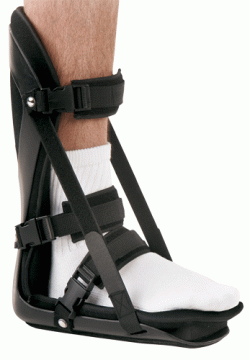
The following self-help treatments have been found to be most effective. Rest your foot. Reduce the amount of weight-bearing activities you participate in. Get off of your feet and elevate them. This will allow healing to begin. Apply ice to your foot. Applications of ice packs that provide a comfortable cooling to the heel and arch (not a freezing cold) will help reduce pain, swelling, and inflammation. Apply the ice to the heel and arch (not the toes). Make sure it is comfortable, and leave on your foot for about 20 minutes, 3 times a day. If you have any medical problems such as diabetes, poor circulation, etc., discuss the use of ice with your doctor before applying the ice. ActiveWrap allows you to apply comfortable cold therapy to your foot without messy ice cubes. Use while on the “go.” Do not walk with bare feet. Always protect your heels, arches, and plantar fascia with good supportive shoes. Orthaheel Orthotic Flip Flops For Men and Women are designed for walking comfort with built in orthotic footbeds that help reduce foot pain from plantar fasciitis. Use in the house or on the beach. Stretch the Plantar Fascia while sleeping. Plantar Fasciitis and Heel Spur pain is usually worse with the first steps in the morning. This is due to the Plantar Fascia tightening up, or contracting while we sleep. To prevent these pain producing contractures of the plantar fascia, the foot must be held in its normal or neutral position while we sleep. This optimal position of the foot is maintained with our comfortable and supportive Night Splint. When foot contractures are prevented during sleep, the “first step pains” Plantar Fasciitis and Heel Spurs will gradually subside. Stretch the Plantar Fascia during the day. Even though the Plantar Fascia is a thick tissue band with very little “give” to it, with the proper care (a Night Splint and the following exercises) it can be stretched a small amount. By stretching the Plantar Fascia even a bit, its abnormal pull on the heel is reduced. This will help to reduce pain and inflammation in the heel and arch. Two of the most effective exercises recommended are. Before stepping down, especially after sleeping or resting, stretch the arch of the foot by stretching your legs out in front of you (do not bend the knee). Place a towel around the ball of the foot. Slowly pull on the ends of the towel, pulling the toes and ball of the foot back as far as is comfortable. Hold the foot in this position for ten seconds. Repeat at least ten times. You should feel a pull on the bottom of the foot, especially in the arch. This stretches the plantar fascia, and reduces its pull on the heel. Stand about 2 to 3 feet from a wall. Lean forward with your hands against the wall. With the painful foot behind, place the other foot forward. Press against the wall, shifting weight over the front foot, while straightening the back leg. Keep the heel of the back foot on the floor and feel the stretch in the heel, Achilles tendon, and calf. Hold this position for ten seconds. Repeat at least ten times, and try to do this three times a day. When these things are achieved, the inflammation and pain of Plantar Fasciitis and Heel Spurs will gradually subside. If you are unsure of the nature of your foot problem, if your pain is intense and does not subside, if you are a diabetic or have other medical problems, if your pain is due to an injury, if an open sore is present, if a mass can be felt, or if you think that you may have an infection, we suggest that before beginning any of the above treatments you consult with your doctor.

Foot Pain


Surgical Treatment

Surgery is not a common treatment for this condition. Approximately 5% of people with plantar fasciitis require surgery if non-surgical methods do not help to relieve pain within a year. The surgical procedure involves making an incision in the plantar fascia in order to decrease the tension of the ligament. Potential risks of this surgical procedure include irritation of the nerves around the heel, continued plantar fasciitis, heel or foot pain, infection, flattening of the arch, problems relating to the anesthetic.


Prevention

Maintain a healthy weight. This minimizes the stress on your plantar fascia. Choose supportive shoes. Avoid high heels. Buy shoes with a low to moderate heel, good arch support and shock absorbency. Don’t go barefoot, especially on hard surfaces. Don’t wear worn-out athletic shoes. Replace your old athletic shoes before they stop supporting and cushioning your feet. If you’re a runner, buy new shoes after about 500 miles of use. Change your sport. Try a low-impact sport, such as swimming or bicycling, instead of walking or jogging. Apply ice. Hold a cloth-covered ice pack over the area of pain for 15 to 20 minutes three or four times a day or after activity. Or try ice massage. Freeze a water-filled paper cup and roll it over the site of discomfort for about five to seven minutes. Regular ice massage can help reduce pain and inflammation. Stretch your arches. Simple home exercises can stretch your plantar fascia, Achilles tendon and calf muscles.